�� ���ђʃ^���p�N���f�[�^�W�iJmol�Łj ��
�� �p��� �b Chime�� �b ���o�����^���^���p�N�� �b �f�[�^�W���j���[ ��
�� GPCR - 2012�N�m�[�x�����w�� �b GPCR�֘A�j���[�X ��
��Jmol�R���e���c���X�g
�s �����\����2at9 �t
���{�f�[�^�W�́CSCOP (Structural Classification of Proteins)��Class: Membrane and cell surface proteins and peptides�Ɏ��^����Ă���f�[�^�̒�����CSOSUI WWW Server�iPSB��SOSUI�𗘗p�������K�Q�Ɓj�Ŗ��ђʗ̈�����Ɛ��肳���f�[�^�𒊏o���Čf�ڂ��C���̖��ђʕ����i��Ƃ��ă�-helix�\���j��I�����ĕ\���̕ύX���ł���悤�ɂ������̂ł��Bh1�`�Ŋe�̈���Ch_all�őS�̈��I�����ĉE���t���[���̃{�^���Ŋe��\���̎w������邱�Ƃ��\�ł��B�ꕔ�̃f�[�^��t1�`�����t_all�Ƃ���̂�TMHMM�ŋ��߂��\���ђʕ����ł��B
�@�Ȃ��C���ђʃ^���p�N���ɂ́C�זE���ɑ��݂����e�̂�C�I���`���l���ȂǁC���ђʕ�������-helix�\���������̂̂ق��ɁC��-sheet�Ō`���������o�����\����L����^���p�N���iPDB�����f�[�^���X�g��1qd5�C1t16_A�Ȃǁj��������܂����CSOSUI�ł̓�-sheet�̈�����o�ł��Ȃ��̂ŁC�����ł͏ȗ����܂��B�ڍׂ͐����i�Ⴆ�C2025�N�Ĕ����\����u�זE�̕��q�����w ��7�Łv [NEW!]�G�\���ɖ��ђʃ^���p�N���摜�j��Web���i�Ⴆ�CMembrane Proteins of Known Structure�Ȃǁj���Q�Ƃ��Ă��������B
1ap9 �qPDB�r�@h1�@h2�@h3�@h4�@h5�@h6�@h7�@h_all�@[Photoreceptor]X-Ray Structure Of Bacteriorhodopsin From Microcrystals Grown In Lipidic Cubic Phases.
1ar1��Chain A �qPDB�r�@h1�@h2�@h3�@h4�@h5�@h6�@h7�@h8�@h9�@h10�@h11�@h12�@h_all�@[Complex (Oxidoreductase/Antibody)]Structure At 2.7 Angstrom Resolution Of The Paracoccus Denitrificans Two-Subunit Cytochrome C Oxidase Complexed With An Antibody Fv Fragment.
1at9 �qPDB�r�@h1�@h2�@h3�@h4�@h5�@h6�@h7�@h_all�@[Photoreceptor]Structure Of Bacteriorhodopsin At 3.0 Angstrom Determined By Electron Crystallography.
2at9 �qPDB�r�@h1�@h2�@h3�@h4�@h5�@h6�@h7�@h_all�@[Photoreceptor]Structure Of Bacteriorhodopsin At 3.0 Angstrom By Electron Crystallography.
1bm1 �qPDB�r�@h1�@h2�@h3�@h4�@h5�@h6�@h7�@h_all�@[Photoreceptor]Crystal Structure Of Bacteriorhodopsin In The Light-Adapted State.
1brd �qPDB�r�@h1�@h2�@h3�@h4�@h5�@h6�@h7�@h_all�@[Photoreceptor]Model for the structure of bacteriorhodopsin based on high-resolution electron cryo-microscopy.
2brd �qPDB�r�@h1�@h2�@h3�@h4�@h5�@h6�@h7�@h_all�@[Photoreceptor]Crystal Structure Of Bacteriorhodopsin In Purple Membrane.
1brr��Chain A �qPDB�r�@h1�@h2�@h3�@h4�@h5�@h6�@h7�@h_all�@[Proton Transport]X-Ray Structure Of The Bacteriorhodopsin Trimer/Lipid Complex.
1brx �qPDB�r�@h1�@h2�@h3�@h4�@h5�@h6�@h7�@h_all�@[Proton Pump]Bacteriorhodopsin/Lipid Complex.
1c3w �qPDB�r�@h1�@h2�@h3�@h4�@h5�@h6�@h7�@h_all�@[Ion Transport]Bacteriorhodopsin/Lipid Complex At 1.55 A Resolution.
1c8r �qPDB�r�@h1�@h2�@h3�@h4�@h5�@h6�@h7�@h_all�@[Ion Transport]Bacteriorhodopsin D96M Br State At 2.0 A Resolution.
1c8s �qPDB�r�@h1�@h2�@h3�@h4�@h5�@h6�@h_all�@[Ion Transport]Bacteriorhodopsin D96N Late M State Intermediate.
1cwq��Chain A �qPDB�r�@h1�@h2�@h3�@h4�@h5�@h6�@h7�@h_all�@[Ion Transport]M Intermediate Structure Of The Wild Type Bacteriorhodopsin In Combination With The Ground State Structure.
1dze* �qPDB�rh1�@h2�@h3�@h4�@h5�@h6�@h7�@h_all�@[Bacteriorhodopsin]Structure Of The M Intermediate Of Bacteriorhodopsin Trapped At 100K.
1e0p��Chain A�i����ԁj* �q�����CPDB�r�@h1�@h2�@h3�@h4�@h5�@h6�@h7�@h_all�@[Photoreceptor]�@���֘A�Q�l���L Intermediate Of Bacteriorhodopsin.
1e12 �qPDB�r�@h1�@h2�@h3�@h4�@h5�@h6�@h7�@h_all�@[Ion Pump]Halorhodopsin, A Light-Driven Chloride Pump.
1ehk��Chain A �qPDB�r�@h1�@h2�@h3�@h4�@h5�@h6�@h7�@h8�@h9�@h10�@h11�@h12�@h13�@h_all�@[Oxidoreductase]Crystal Structure Of The Aberrant Ba3-Cytochrome-C Oxidase From Thermus Thermophilus.
1eul �qPDB�r h1�@h2�@h3�@h4�@h5�@h6�@h7�@h8�@h9�@h_all�@[Hydrolase | Ca�|���v]Crystal Structure Of Calcium ATPase With Two Bound Calcium Ions.
1f4z �qPDB�r�@h1�@h2�@h3�@h4�@h5�@h6�@h7�@h_all�@[Proton Transport, Membrane Protein]Bacteriorhodopsin-M Photointermediate State Of The E204Q Mutant At 1.8 Angstrom Resolution.
1f50 �qPDB�r�@h1�@h2�@h3�@h4�@h5�@h6�@h7�@h_all�@[Proton Transport, Membrane Protein]Bacteriorhodopsin-Br State Of The E204Q Mutant At 1.7 Angstrom Resolution.
1f88��Chain A �q�����CPDB�rh1�@h2�@h3�@h4�@h5�@h6�@h7�@h_all �b t1�@t2�@t3�@t4�@t5�@t6�@t7�@t_all�@[Signaling Protein]Crystal Structure Of Bovine Rhodopsin.
1fbb �qPDB�r�@h1�@h2�@h3�@h4�@h5�@h6�@h7�@h_all�@[Proton Transport]Crystal Structure Of Native Conformation Of Bacteriorhodopsin.
1fbk �qPDB�r�@h1�@h2�@h3�@h4�@h5�@h6�@h7�@h_all�@[Proton Transport]Crystal Structure Of Cytoplasmically Open Conformation Of Bacteriorhodopsin.
1fft��Chain A �qPDB�r�@h2�ih1�Ȃ��j�@h3�@h4�@h5�@h6�@h7�@h8�@h9�@h10�@h11�@h12�@h13�ih14-15�Ȃ��j�@h_all�ih2-13�j�@[Oxidoreductase]The Structure Of Ubiquinol Oxidase From Escherichia Coli.
1fqy �qPDB�r h1�@h2�@h3�@h4�@h5�@h_all�@[Membrane Protein | 2003�N�m�[�x�����w�� | �A�N�A�|����]Structure Of Aquaporin-1 At 3.8 �� Resolution By Electron Crystallography.
1fx8 �qPDB�r�@h1�@h2�@h3�@h4�@h5�@h6�@h_all�@[Membrane Protein]Crystal Structure Of The E. Coli Glycerol Facilitator (Glpf) With Substrate Glycerol.
1gu8 �qPDB�r�@h1�@h2�@h3�@h4�@h5�@h6�@h7�@h_all�@[Photoreceptor]
1gue �qPDB�r�@h1�@h2�@h3�@h4�@h5�@h6�@h7�@h_all�@[Photoreceptor]
1h2s��Chain A �qPDB�r�@h1�@h2�@h3�@h4�@h5�@h6�@h7�@h_all�@[Menbrane Protein Complex]Molecular Basis Of Transmenbrane Signalling By Sensory Rhodopsin �U-Transducer Complex.
1h68 �qPDB�r�@h1�@h2�@h3�@h4�@h5�@h6�@h7�@h_all�@[Photoreceptor]
1h6i �qPDB�r�@h1�@h2�@h3�@h4�@h5�@h_all�@[Membrane Protein]A Refined Structure Of Human Aquaporin 1.
1hzx��Chain A�qPDB�r�@h1�@h2�@h3�@h4�@h5�@h6�@h7�@h_all�@[Signaling Protein]Crystal Structure Of Bovine Rhodopsin.
1ih5 �qPDB�r�@h1�@h2�@h3�@h4�@h5�@h_all�@[Membrane Protein]Crystal Structure Of Aquaporin-1.
1iw6 �qPDB�r�@h1�@h2�@h3�@h4�@h5�@h6�@h7�@h_all�@[Proton Transport]Crystal Structure Of The Ground State Of Bacteriorhodopsin.
1iwo��Chain A �qPDB�CeProtS�rh1�@h2�@h3�@h4�@h5�@h6�@h7�@h8�@h9�@h_all�@ [Hydrolase | �J���V�E��ATP�A�[�[]Crystal Structure Of The Sr Ca2+-ATPase In The Absence Of Ca2+.
1ixf �qPDB�r�@h1�@h2�@h3�@h4�@h5�@h6�@h7�@h_all�@[Proton Transport]Crystal Structure Of The K Intermediate Of Bacteriorhodopsin.
1j4n*�i�`���l�����j �qPDB�rh1�@h2�@h3�@h4�@h5�@h_all �b t1�@t2�@t3�@t4�@t5�@t6�@t_all�@[Membrane Protein | �A�N�A�|����]Crystal Structure Of The Aqp1 Water Channel.
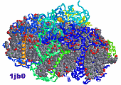
1jb0��Chain A �qPDB�r�@h1�@h2�@h3�@h4�@h5�@h6�@h7�@h_all�@[Photosynthesis]Crystal Structure Of Photosystem I: A Photosynthetic Reaction Center and Core Antenna System From Cyanobacteria.
 ��1jb0�̑S�̍\�� ��1jb0�̑S�̍\��
1jfp �qPDB�rh1�@h2�@h3�@h4�@h5�@h6�@h7�@h_all�@[Signaling Protein, Membrane Protein]Structure Of Bovine Rhodopsin (Dark Adapted).
1jgj* �q�����CPDB�r�@h1�@h2�@h3�@h4�@h5�@h6�@h7�@h_all�@[Signaling Protein]Crystal Structure Of Sensory Rhodopsin �U At 2.4 Angstroms: Insights Into Color Tuning and Transducer Interaction.
1jv6 �qPDB�r�@h1�@h2�@h3�@h4�@h5�@h6�@h7�@h_all�@[Ion Transport]Bacteriorhodopsin D85S/F219L Double Mutant At 2.00 Angstrom Resolution.
1jv7 �qPDB�r�@h1�@h2�@h3�@h4�@h5�@h6�@h7�@h_all�@[Ion Transport]Bacteriorhodopsin O-Like Intermediate State Of The D85S Mutant At 2.25 Angstrom Resolution.
1kg8 �qPDB�r�@h1�@h2�@h3�@h4�@h5�@h6�@h7�@h_all�@[Proton Transport]X-Ray Structure Of An Early-M Intermediate Of Bacteriorhodopsin.
1kg9 �qPDB�r�@h1�@h2�@h3�@h4�@h5�@h6�@h7�@h_all�@[Proton Transport]Structure Of A "Mock-Trapped" Early-M Intermediate Of Bacteriorhosopsin.
1kgb �qPDB�r�@h1�@h2�@h3�@h4�@h5�@h6�@h7�@h_all�@[Lyase (Aldehyde) Proton Transport]Structure Of Ground-State Bacteriorhodopsin.
1kju �qPDB�r�@h1�@h2�@h3�@h4�@h5�@h6�@h7�@h8�@h9�@h_all�@[Hydrolase]Ca2+-ATPase In The E2 State.
1kme��Chain A �qPDB�r�@h1�@h2�@h3�@h4�@h5�@h6�@h7�@h_all�@[Membrane Protein]Crystal Structure Of Bacteriorhodopsin Crystallized From Bicelles.
1kpk��Chain A �qPDB�r�@h1�@h2�@h3�@h4�@h5�@h6�@h7�@h8�@h9�@h10�@h_all�@[Membrane Protein]Crystal Structure Of The Clc Chloride Channel From E. Coli.
1kpl��Chain A �qPDB�r�@h1�@h2�@h3�@h4�@h5�@h6�@h7�@h8�@h9�@h10�@h_all�@[Membrane Protein]Crystal Structure Of The Clc Chloride Channel From S. Typhimurium.
1l0m �qPDB�r�@h1�@h2�@h3�@h4�@h5�@h6�@h_all�@[Proton Transport]Solution Structure Of Bacteriorhodopsin.
1l7v��Chain A �qPDB�r�@h1�@h2�@h3�@h4�@h5�@h6�@h7�@h8�@h_all�@[Transport Protein/Hydrolase]Bacterial Abc Transporter Involved In B12 Uptake.
1l9h��Chain A �qPDB�r�@h1�@h2�@h3�@h4�@h5�@h6�@h7�@h_all�@[Signaling Protein]Crystal Structure Of Bovine Rhodopsin At 2.6 Angstroms Resolution.
1lda �qPDB�r�@h1�@h2�@h3�@h4�@h5�@h6�@h_all�@[Transport Protein]Crystal Structure Of The E. Coli Glycerol Facilitator (Glpf) Without Substrate Glycerol.
1ldf �qPDB�r�@h1�@h2�@h3�@h4�@h5�@h6�@h_all�@[Transport Protein]Crystal Structure Of The E. Coli Glycerol Facilitator (Glpf) Mutation W48F, F200T.
1ldi �qPDB�r�@h1�@h2�@h3�@h4�@h5�@h6�@h_all�@[Transport Protein]Crystal Structure Of The E. Coli Glycerol Facilitator (Glpf) Without Substrate Glycerol.
1ln6 �qPDB�r�@h1�@h2�@h3�@h4�@h5�@h6�@h7�@h_all�@[Membrane Protein]Structure Of Bovine Rhodopsin (Metarhodopsin �U).
1m0k �qPDB�r�@h1�@h2�@h3�@h4�@h5�@h6�@h7�@h_all�@[Ion Transport]Bacteriorhodopsin K Intermediate At 1.43 A Resolution.
1m0l �qPDB�r�@h1�@h2�@h3�@h4�@h5�@h6�@h7�@h_all�@[Ion Transport]Bacteriorhodopsin/Lipid Complex At 1.47 A Resolution.
1m0m �qPDB�r�@h1�@h2�@h3�@h4�@h5�@h6�@h7�@h_all�@[Ion Transport]Bacteriorhodopsin M1 Intermediate At 1.43 A Resolution.
1m56��Chain A �qPDB�r�@h1�@h2�@h3�@h4�@h5�@h6�@h7�@h8�@h9�@h10�@h11�@h12�@h_all�@[Oxidoreductase]Structure Of Cytochrome C Oxidase From Rhodobactor Sphaeroides (Wild Type).
1m57��Chain A �qPDB�r�@h1�@h2�@h3�@h4�@h5�@h6�@h7�@h8�@h9�@h10�@h11�@h12�@h_all�@[Oxidoreductase]Structure Of Cytochrome C Oxidase From Rhodobacter Sphaeroides (Eq(I-286) Mutant)).
1mgy �qPDB�r�@h1�@h2�@h3�@h4�@h5�@h6�@h7�@h_all�@[Proton Transport]Structure Of The D85S Mutant Of Bacteriorhodopsin With Bromide Bound.
1o0a �qPDB�r�@h1�@h2�@h3�@h4�@h5�@h6�@h7�@h_all�@[Proton Transport]Bacteriorhodopsin L Intermediate At 1.62 A Resolution.
1occ��Chain A �qPDB�r�@h1�@h2�@h3�@h4�@h5�@h6�@h7�@h8�@h9�@h10�@h11�@h12�@h_all�@[Oxidoreductase (Cytochrome(C)-Oxygen)]Structure Of Bovine Heart Cytochrome C Oxidase At The Fully Oxidized State.
2occ��Chain A �qPDB�r�@h1�@h2�@h3�@h4�@h5�@h6�@h7�@h8�@h9�@h10�@h11�@h12�@h_all�@[Oxidoreductase]Bovine Heart Cytochrome C Oxidase At The Fully Oxidized State.
1oco��Chain A �qPDB�r�@h1�@h2�@h3�@h4�@h5�@h6�@h7�@h8�@h9�@h10�@h11�@h12�@h_all�@[Oxidoreductase]Bovine Heart Cytochrome C Oxidase In Carbon Monoxide-Bound State.
1ocr��Chain A �qPDB�r�@h1�@h2�@h3�@h4�@h5�@h6�@h7�@h8�@h9�@h10�@h11�@h12�@h_all�@[Oxidoreductase]Bovine Heart Cytochrome C Oxidase In The Fully Reduced State.
1ocz��Chain A �qPDB�r�@h1�@h2�@h3�@h4�@h5�@h6�@h7�@h8�@h9�@h10�@h11�@h12�@h_all�@[Oxidoreductase]Bovine Heart Cytochrome C Oxidase In Azide-Bound State.
1ots��Chain A �qPDB�r�@h1�@h2�@h3�@h4�@h5�@h6�@h7�@h8�@h9�@h10�@h_all�@[Membrane Protein]Structure Of The Escherichia Coli Clc Chloride Channel and Fab Complex.
1ott��Chain A �qPDB�r�@h1�@h2�@h3�@h4�@h5�@h6�@h7�@h8�@h9�@h10�@h_all�@[Membrane Protein]Structure Of The Escherichia Coli Clc Chloride Channel E148A Mutant and Fab Complex.
1otu��Chain A �qPDB�r�@h1�@h2�@h3�@h4�@h5�@h6�@h7�@h8�@h9�@h10�@h_all�@[Membrane Protein]Structure Of The Escherichia Coli Clc Chloride Channel E148Q Mutant and Fab Complex.
1oy8 �qPDB�r�@h1�@h2�@h3�@h4�@h5�@h6�@h7�@h8�@h9�@h10�@h11�@h_all�@[Membrane Protein]Structural Basis Of Multiple Drug Binding Capacity Of The Acrb Multidrug Efflux Pump.
1oy9 �qPDB�r�@h1�@h2�@h3�@h4�@h5�@h6�@h7�@h8�@h9�@h10�@h11�@h_all�@[Membrane Protein]Structural Basis Of Multiple Drug Binding Capacity Of The Acrb Multidrug Efflux Pump.
1oyd �qPDB�r�@h1�@h2�@h3�@h4�@h5�@h6�@h7�@h8�@h9�@h10�@h11�@h_all�@[Membrane Protein]Structural Basis Of Multiple Binding Capacity Of The Acrb Multidrug Efflux Pump.
1oye �qPDB�r�@h1�@h2�@h3�@h4�@h5�@h6�@h7�@h8�@h9�@h10�@h11�@h_all�@[Membrane Protein]Structural Basis Of Multiple Binding Capacity Of The Acrb Multidrug Efflux Pump.
1p8h �qPDB�r�@h1�@h2�@h3�@h4�@h5�@h6�@h7�@h_all�@[Proton Transport]Bacteriorhodopsin M1 Intermediate Produced At Room Temperature.
1p8i �qPDB�r�@h1�@h2�@h3�@h4�@h5�@h6�@h7�@h_all�@[Proton Transport]F219L Bacteriorhodopsin Mutant.
1p8u �qPDB�r�@h1�@h2�@h3�@h4�@h5�@h6�@h7�@h_all�@[Proton Transport]Bacteriorhodopsin N' Intermediate At 1.62 A Resolution.
1pf4��Chain A�EB �qPDB�r�@h1�@h2�@h3�@h4�@h5�@h6�@h_all�@[Lipid Transport]Structure Of Msba From Vibrio Cholera: A Multidrug Resistance Abc Transporter Homolog In A Closed Conformation.
1qhj �qPDB�r�@h1�@h2�@h3�@h4�@h5�@h6�@h7�@h_all�@[Photoreceptor]X-Ray Structure Of Bacteriorhodopsin Grown In Lipidic Cubic Phases.
1qko �qPDB�r�@h1�@h2�@h3�@h4�@h5�@h6�@h7�@h_all�@[Photoreceptor]High Resolution X-Ray Structure Of An Early Intermediate In The Bacteriorhodopsin Photocycle.
1qkp �qPDB�r�@h1�@h2�@h3�@h4�@h5�@h6�@h7�@h_all�@[Photoreceptor]High Resolution X-Ray Structure Of An Early Intermediate In The Bacteriorhodopsin Photocycle.
1qle��Chain A �qPDB�r�@h1�@h2�@h3�@h4�@h5�@h6�@h7�@h8�@h9�@h10�@h11�@h12�@h_all�@[Complex (Oxidoreductase/Antibody)]Cryo-Structure Of The Paracoccus Denitrificans Four-Subunit Cytochrome C Oxidase In The Completely Oxidized State Complexed With An Antibody Fv Fragment.
1qm8 �qPDB�r�@h1�@h2�@h3�@h4�@h5�@h6�@h7�@h_all�@[Photoreceptor]Structure Of Bacteriorhodopsin At 100 K.
1uaz��Chain A �qPDB�r�@h1�@h2�@h3�@h4�@h5�@h6�@h7�@h_all�@[Proton Transport]Crystal Structure Of Archaerhodopsin-1.
|

